Temporomandibular Joint Dysfunction

What is the Temporomandibular Joint? 

It is a joint which connects the jaw bone with the base of the skull. It has a hinge-shaped action, which allows the mouth to be opened and closed just in front of the ear (See Image).

What is the Temporomandibular Joint Dysfunction?

Currently, there is scientific evidence which shows that this condition is more common in female patients between the ages of 20 and 40. It manifests itself when there is inflammation or wear of the structures within the joint which limits or impairs the opening and closing of the mouth when speaking, yawning, chewing or swallowing. Temporomandibular joint dysfunction can affect one or both sides, as well as nearby muscles, causing pain, tension, and inflammation. 

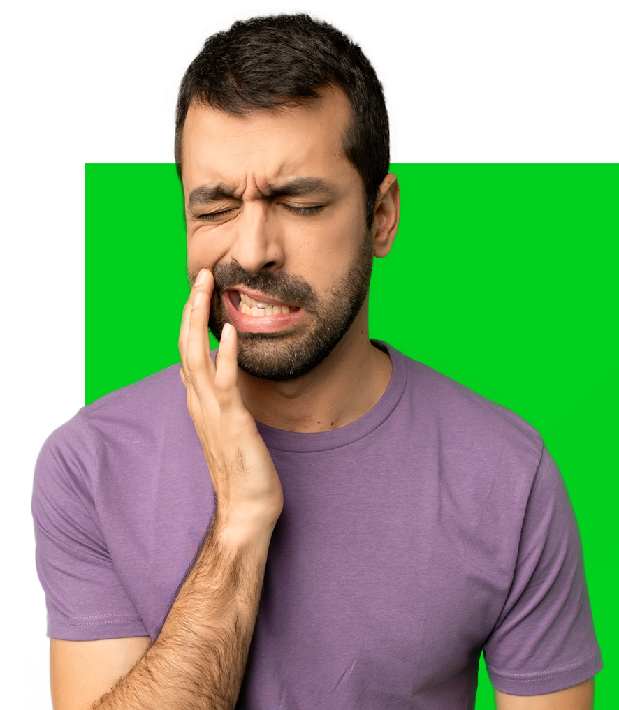
The most common cause of this condition is constant grinding, gnashing, or clenching of the teeth (also known as bruxism). Bruxism may happen unconsciously, when you are awake, or when you sleep. The constant movement of the joint not only inflames or erodes the structures of the joint, it also produces a noticeable wear of the teeth (see image).


Salud, O. (2021, January 5). What is bruxism? Symptoms and treatment. https://odontologiasalud.blogspot.com/2009/03/que-es-el-bruxismo.html

What are the symptoms?

The most common symptoms are:

  • Pain in the affected jaw or in the joint area (just in front of the ear) at rest or movement (eating, speaking, yawning, etc). 
  • Pain on one side of the face or in the jaw in front of the ear or on the affected side. 
  • Headache (particularly in the morning)
  • Pain or limitation when opening the mouth. 
  • Ear pain
  • 'Click' when opening and closing the mouth. 
  • Pain on one side of the face or neck. 
  • Feeling of blocked ear or dizziness. 

How is Temporomandibular Joint Dysfunction diagnosed and treated?

It is important to see a specialist (otolaryngologist) when having discomfort or pain in the jaw or in the joint area (just in front of the ear) at rest or movement (eating, speaking, yawning, etc.), which it does not improve with conventional analgesic treatment. 

During the consultation, the specialist doctor will not only take note of your important medical history, he will also examine your ear and jaw region in order to diagnose the cause of your condition. It is important to emphasize the importance of going to a specialist doctor since due to pain in the ear region caused by an inflamed temporomandibular joint, it is common for it to be misdiagnosed and treated as an ear infection. 

Treatment can vary depending on the severity of symptoms and damage to the temporomandibular joint. It is advisable to start with conservative treatment:

  • Application of local heat with a towel moistened in warm / hot water and massage in the affected temporomandibular region several times a day.
  • Avoid hard foods that put additional pressure on the joint, opt for foods with a soft consistency. 
  • Restrict the opening of the mouth when biting food, chewing, talking or yawning.
  • Reduce stress (meditation, yoga, deep breaths, improve sleep habits, aerobic exercise, etc.), as well as be aware of when we clench or grind our teeth and avoid it whenever possible. 
  • In severe cases, it will also be necessary to take anti-inflammatory drugs with or without a muscle relaxant.

Your doctor may also assess the need to treat your case together with a:

  • Psychologist or psychiatrist to improve stress or anxiety management. 
  • Dentist to assess the condition of your teeth and evaluate the use of a dental guard to control involuntary movements of the teeth during sleep. 

If you have noticeable improvement from medical treatment, your doctor may suggest surgical treatment. There are several techniques and they vary depending on the cause and severity of the temporomandibular dysfunction; your doctor will recommend the best treatment for you. 

The decision to have any surgical procedure should be after the surgeon knows all the patient’s medical history and has fully explained as well as discussed the surgical procedure, possible complications and risks.

* This information does not take the place of your doctor's advice. Please consult your healthcare provider for information about a specific medical condition.

References

Cope, G., & Cope, A. (2011). Diagnosis, treatment and management of TMJ disorders. Dental Nursing, 7(12), 682–686. https://doi.org/10.12968/denn.2011.7.12.682 

Flint, P. W., Flint, P. W., & Cummings, C. W. (2020). Cummings otolaryngology: head and neck surgery; Chapter 89: Temporomandibular Joint Disorders (7th ed., Vol. 2). Elsevier. 

What is TMJ? Mountain Ear Nose and Throat Associates. (n.d.). https://mountainent.com/blog/what-is-tmj. 

Turbinates and nasal obstruction

What are turbinates?

The turbinates are structures on the sides of the nose responsible for directing, filtering and humidifying the air we breathe through the nose (see image). We generally have three pairs in each nostril, named for their location: inferior turbinates, middle turbinates, and superior turbinates. 

Each turbinate is made up of bone surrounded by mucosa, which is lined with blood vessels and nerve endings. In turn, the mucosa surrounding the turbinates contain immune receptors, which help to perceive and block the inhalation of irritants or possible causes of infection.

http://www.beverlyhillssinus.com/nasal-sinus-surgery/nasal-turbinate-reduction/.

Can turbinates cause nasal obstruction?

The growth or inflammation of the turbinates may prevent you from breathing properly through one or both nostrils. The turbinates can become inflamed by exposure to an allergic agent (such as dust, pollen, mites, etc.), by inhaling irritants, changes in temperature, or due to infection in the nose or sinuses. Most of these cases are temporary conditions, and the turbinates return to their normal size.

However, continued exposure to irritants or allergens can cause persistent inflammation of the turbinates, particularly the lower turbinates (see image). This, depending on the severity, can cause us to breathe through the mouth most of the time which can limit physical activity, cause bad breath, hoarseness or sleep-related breathing disorders, headaches, runny nose and, in the childhood can affect the development of certain structures in the face.

Turbinate Reduction St. Louis, MO: Turbinate Reduction Surgery. St. Louis Sinus Center. (2020, December 3). https://www.stlsinuscenter.com/common-sinus-problems/inferior-turbinate-hypertrophy/turbinate-reduction-surgery/.

How is persistent turbinate inflammation diagnosed and treated?

It is important to see a specialist in the nose and paranasal sinuses (otolaryngologist) if you suspect or have a persistent nasal obstruction for more than three weeks. 

During the consultation, the specialist doctor will not only take note of your important medical history, he will also examine your nose in order to diagnose the cause of your condition either manually or with the help of a small camera called an endoscope. 

Initial treatment for inflamed turbinates can vary depending on the doctor or medical condition and is focused on reducing inflammation and improving nose breathing. Anti-inflammatories or antihistamines are usually prescribed either as a spray applied directly to the nose or orally. 

If you do not have noticeable improvement with medical treatment, your doctor may suggest a simple surgical procedure to reduce the size of the turbinates called a turbinoplasty. There are several surgical techniques to reduce the size of the turbinates; your doctor will recommend the best treatment for you. In general, most procedures do not require incisions in the skin, and do not change the shape or size of the nose. 

The decision to have any surgical procedure should be after the surgeon knows all the patient’s medical history and has fully explained as well as discussed the surgical procedure, possible complications and risks.

* This information does not take the place of your doctor's advice. Please consult your healthcare provider for information about a specific medical condition.

References

Abdullah, B., & Singh, S. (2021). Surgical Interventions for Inferior Turbinate Hypertrophy: A Comprehensive Review of Current Techniques and Technologies. International Journal of Environmental Research and Public Health, 18(7), 3441.

https://doi.org/10.3390/ijerph18073441 Becker, D. (2003). Septoplasty and turbinate surgery. Aesthetic Surgery Journal, 23(5), 393–403. https://doi.org/10.1016/s1090-820x(03)00208-5

Septum or Nasal Septum

What is the septum or nasal septum?

The septum or nasal septum is located in the middle of the two nostrils or nostrils; It is lined by mucosa which produces mucus, and is made up of cartilage and bone. This structure divides the nose in two, directs the air we breathe into the airway, and provides support and structure to the nose (see image). 

Source: U.S. National Library of Medicine. (n.d.). Septoplastia – Serie-Indicaciones: MedlinePlus enciclopedia médica. MedlinePlus. https://medlineplus.gov/spanish/ency/esp_presentations/100038_2.htm.

What causes a deviated septum and what are the symptoms?

The septum can be twisted or deviated as we grow during childhood, or when we suffer a blow to the nose (see image).

Source: U.S. National Library of Medicine. (n.d.). Septoplastia – Serie-Indicaciones: MedlinePlus enciclopedia médica. MedlinePlus. https://medlineplus.gov/spanish/ency/esp_presentations/100038_2.htm.

The most common symptom is the inability to breathe properly through one or both nostrils. This, depending on the severity, can cause a deformity in the nose or that we breathe through the mouth most of the time which can limit physical activity, cause bad breath, hoarseness or sleep-related breathing disorders, recurrent nosebleeds, headaches, runny nose and, in childhood it can affect the development of certain structures in the face.


In severe cases the deformity of the nasal septum can obstruct the drainage of the paranasal sinuses causing recurrent infections or sinusitis (click here to read what it is sinusitis), alterations in smell, pressure or pain in the face, fever and a feeling of blocked ears.

How is a septal deviation diagnosed and how is it corrected?

It is important to see a specialist in the nose and paranasal sinuses (otolaryngologist) if you suspect or have symptoms caused by a deviated nasal septum. 

During the consultation, the specialist doctor will not only take note of your important medical history, he will also examine your nose in order to diagnose the cause of your condition either manually or with the help of a small camera called an endoscope. 

En caso de tener una desviación septal, el médico podrá solicitar una tomografía computarizada de nariz y senos paranasales, el cual brinda imágenes detalladas del interior de tu nariz y senos paranasales para lograr visualizar detalladamente el grado  y localización de desviación del septum, o si existen algún otra condición médica asociada (ver imagen).

CT scan showing nasal septum deviation (yellow arrow)

In turn, if surgical treatment is required, this study also helps the surgeon to plan the surgery in order to provide you with the best possible results. 

By going to an otolaryngologist, you will be given a diagnosis and treatment. Commonly a deviation of the nasal septum which causes symptoms and does not improve with medical treatment, requires a surgical correction called rhinoseptumplastia or septumplastia. There are several surgical techniques to correct a septal deviation, such as: closed, open or endoscopic approach. Your doctor will recommend the best treatment for you. 

The decision to have any surgical procedure should be after the surgeon knows all the patient’s medical history and has fully explained as well as discussed the surgical procedure, possible complications and risks.

* This information does not take the place of your doctor's advice. Please consult your healthcare provider for information about a specific medical condition.

References 

Becker, D. (2003). Septoplasty and turbinate surgery. Aesthetic Surgery Journal, 23(5), 393–403. https://doi.org/10.1016/s1090-820x(03)00208-5 

Grover, N. (2017). Septoplasty and Nasal Airway Surgery. Otorhinolaryngology: Head and Neck Surgery (2 Volumes), 1894–1894. https://doi.org/10.5005/jp/books/13036_130 

Nedev, P. (2010). Rhinoseptoplasty – complications, mistakes or unpredictable deformities. International Bulletin of Otorhinolaryngology, 6(1), 23. https://doi.org/10.14748/orl.v6i1.7058 

U.S. National Library of Medicine. (n.d.). Septoplastia – Serie-Indicaciones: MedlinePlus enciclopedia médica. MedlinePlus. https://medlineplus.gov/spanish/ency/esp_presentations/100038_2.htm. 

Cerumen (Earwax) Impaction

¿What is earwax?

Cerumen, commonly called earwax, is produced by small glands located at the outer part of our external ear canal. Cerumen not only protects the skin in our ear external canal from infection by reducing moisture and creating an acidic environment. It also prevents foreign bodies (like insects) or dust from entering the ear canal, and reduces the exposure to loud noises that could potentially damage the inner ear.

¿How does Cerumen get impacted?

As cerumen is produced it traps old skin and/or dust in our externa ear canal, and with jaw movements and chewing motions it is naturally moved towards the entrance of the ear. In normal circumstances simply cleaning the excess cerumen from the outer area of your ear is enough and should be all the cleaning you need to do.

However, this natural cleansing mechanism can be affected by many health conditions.

A few examples are:

⦁ A reduced diameter of the external ear canal: some people have naturally a narrower/small ear canal or excess hair growth which can cause the earwax to build up. However, this can also be caused by ear infections, genetic malformations of the skull or ear canal (such as Down Syndrome), bony growths inside of the ear canal and non-cancer or cancer tissue growth.

⦁ Excess or dense production of ear wax: some people produce an excessive amount of earwax, and as we age the glands in our ear canal produce a more dense/think earwax, which over exceeds or interrupts our natural cleansing mechanisms. Also, ear wax can be over produced as a response to a foreign body or trauma such as over use of self-cleansing objects such as Q-tips or overuse of headphones.

⦁ Overuse of cotton swabs or Q-Tips: use of cotton swabs seem harmful if they are used to clean the out part of the ear, without introducing it into the ear canal. When introduced deep into the ear canal it not only stimulates your body to produce more ear wax, it also pushes the ear wax further in, creating a more dense and hard to remove plug.

source: https://dizziness-and-balance.com/disorders/unilat/otitis.html
source: Tomar, M., Devi, H.P., Khundrakpam, R., Pant, A., & Saha, R. (2018). Study for evaluation of etiology , clinical presentation , audiometric assessment and spontaneous healing of traumatic perforation of tympanic membrane.

How do I know I have ear way accumulation or impaction?

Ear wax accumulation is usually asymptomatic. However, a few patients can suffer from:

⦁ Sudden or gradual hearing loss.

⦁ Ear pain

⦁ Blocked/itchy ears

⦁ Dizziness

⦁ Cough

How can I prevent earwax accumulation or impaction?

⦁ Avoid introducing any object into your ear canal. I always tell my patients, “the ear should be cleaned with your elbows”.

⦁ Try to “soften” the earwax: Use a baby dropper or eye dropper to apply 2- 3 drops of baby oil, mineral oil or liquid Vaseline once a day into your ear canal.

⦁ Use warm water to help the natural ear wax cleaning process: while taking a shower gently aim the stream of the shower head into your ear canal (avoid strong water jet streams) this will soften the ear wax and help remove any excess in the outer part of your ear.

⦁ Dry your ear canal: you can do this by simply aiming a hair drying with the cold setting (avoid hot air) into your ear canal for a few minutes after shower or swimming.

⦁ Visit your favorite ear nose and throat specialist: if there is any discomfort in your ears, or you know you tend to suffer from earwax impaction, it is recommended to have your ears check by a specialist every 6 months or when then symptoms occur.

* This information does not take the place of your doctor's advice. Please consult your healthcare provider for information about a specific medical condition.

References

Guest, J., Greener, M., Robinson, A., & Smith, A. (2004). Impacted cerumen: Composition, production, epidemiology and management. QJM, 97(8), 477-488. doi:10.1093/qjmed/hch082

Seth et al. (2017). Clinical Practice Guideline (Update): Earwax (Cerumen Impaction). Head and Neck Surgery, Vol. 156(1S) S1–S29


Tomar, M., Devi, H.P., Khundrakpam, R., Pant, A., & Saha, R. (2018). Study for evaluation of etiology , clinical presentation , audiometric assessment and spontaneous healing of traumatic perforation of tympanic membrane.

Noise Induced Hearing Loss

What are the mayor structures of the ear?

The ear is divided in three main parts (see image below):

the external ear: formed by the ear lobe, the external ear canal and the ear drum or tympanic membrane.

the middle ear: includes the small bones (ossicles) called malleus, incus and stapes. The middle ear is ventilated and inner pressure is equalized via a narrow tube located at the back of the nose called the Eustachian tube.

the inner ear: includes a spiral-shaped structure called cochlea. The cochlea has small hair like structures called stereocilia which perceive sound waves and transduce them into electrical impulses that can be interpreted by the brain.

Source: Kiversal. (2019, December). The ear: structure and functions. Blog of Kiversal. https://blog.kiversal.com/en/the-ear-structure-and-functions/.

How do we perceive sound?

Sound waves hit your ear lobe, channels down to your external ear canal which makes the ear drum vibrate. This vibration jiggles the small bones in the middle ear. Then, the vibration is passed into the cochlea which stimulate the sterocilla, creating electrical impulses which are then transmitted and perceived as sound in your brain.

What causes hearing loss?

Two of the most common causes of hearing loss in adults are due to the natural aging process, also called presbycusis, and noise induced hearing loss. Every time you are exposed to a loud sound you have the potential risk of damaging your hearing. An excessive amount of sound, causes intense stimulation to the small hair like structures in your inner ear. These intense vibrations can cause permanent damage: meaning not reversible.

Two of the most common causes of hearing loss in adults are due to the natural aging process, also called presbycusis, and noise induced hearing loss. Every time you are exposed to a loud sound you have the potential risk of damaging your hearing. An excessive amount of sound, causes intense stimulation to the small hair like structures in your inner ear. These intense vibrations can cause permanent damage: meaning not reversible. According to the World Health Organization, there are approximately 1.1 billion young individuals at risk of permanently damaging their hearing due to exposure of unsafe levels of noise which includes: bars, nightclubs, cinemas, concerts, live events, fitness classes and listening to music or video game consoles over personal audio devices.

Individuals exposed to constant loud noises are three times likely to irreversibly lose their hearing at an early age. Early loss of hearing is a direct cause of depression, personal isolation, inability to communicate, inefficient job performance and frustration.

There are many other causes of hearing loss including head injury, illness and certain medication that can be toxic for the inner ear.

There are more than 200 medications on the market that are known to be toxic to the inner ear. Risk of ototoxicity depends on the way of administering the medication (intravenously has the highest risk), dose, age, and general health condition. You should ask your doctor if you are at risk before taking any medication. Just a few examples are:

⦁ Non-Steroidal Anti-Inflammatory Drugs (NSAIDs), such as aspirin, ibuprofen, or naproxen.

⦁ Certain antibiotics, including aminoglycosides.

⦁ Certain chemotherapy drugs for cancer treatment.

⦁ Blood pressure pills and diuretics.

⦁ Quinine-based medications (Chloroquine, Quinacrine Hydrochloride, Quinine Sulfate).

How to prevent can early hearing loss?

The number one thing you can do to keep your hearing as healthy as possible is to avoid or limit exposure to loud noises. Some of the common sources are:

⦁ Loud music (concerts, live events, fitness classes)

⦁ Heavy machinery

⦁ Gun fire

⦁ Excessive use of head phones for prolonged periods of time.

Noise is measured with a complex mathematic algorithm called decibels: in general terms the higher the decibels the stronger the wave sound and impact in our ear.

The World health Organization main recommendations are:

⦁ Exposure to 85 dB for a maximum limit of eight hours per day.

⦁ Followed by at least ten hours of recovery time at 70 dBA or lower.

⦁ Noise levels above 140 dB are not considered safe.

See the image below to learn the decibel levels of common everyday sounds:

Source: A Complete Guide to Noise-Induced Hearing Loss. EarQ. (2020, April). https://www.earq.com/hearing-health/articles/guide-to-noise-induced-hearing-loss.

This does not mean you should avoid listening to music or going concerts. The best way to prevent any hearing damage is to be aware of the noise level and how long you are exposed to it. Remember, once it passes a certain level, then it can permanently damage your hearing.

How can I prevent Noise induced hearing loss?

⦁ Wear earplugs: conventional ear plugs can reduce noise by 20-30 decibels. If your job involves loud noises there are high fidelity earplugs called `musicians earplugs which reduce noise levels while maintaining the quality of the original sound.

⦁ Respect the 60/60 rule: When listening to music with a personal audio device, limit the sound to 60% volume for no more than 60 minutes a day. Currently, most smartphones allow you to set a limit on the sound volume. So, limiting sound to a maximum of 60% can prevent any permanent hearing damage in the future.

⦁ Use over-the-ear headphones: devices that sit over your ears are safer since they are further away from the structures inside your ear.

⦁ Download smartphone apps: There are many smartphone apps that monitor the volume level of your smartphone or your surroundings. This can increase awareness and warn you if you are exposed to dangerous noise levels.

⦁ Take a break and recover: When exposed to loud noises at concerts, nightclubs or live events give your ears time to recover, and take breaks from the loud noise. Also, after a night out to a nightclub or after a concert, take at least 10 hours of ‘quiet’ to give your ears time to recover from the stress.

Remember noise induced hearing loss is preventable!

If you take care of the amount of sound you are exposed to and use effective prevention measures you will be able to enjoy sounds for a long time!

* This information does not take the place of your doctor's advice. Please consult your healthcare provider for information about a specific medical condition.

References

A Complete Guide to Noise-Induced Hearing Loss. EarQ. (2020, April). https://www.earq.com/hearing-health/articles/guide-to-noise-induced-hearing-loss.

Banzet, E. (2019, May). Noise-Induced Hearing Loss. speechandhearing.ca / paroleetaudition.ca. https://speechandhearing.ca/noise-induced-hearing-loss/.

Flint, P. W., & Cummings, C. W. (2021). Cummings Otolaryngology: head and neck surgery -Noise-Induced Hearing Loss. In Cummings Otolaryngology: head and neck surgery (pp.2140-2152). essay, Elsevier.

Noise Induced Hearing Loss among University Students Using Portable Entertainment Player. (2018). International Journal of Otorhinolaryngology, 5(1), 01–02. https://doi.org/10.13188/2380-0569.1000025

Prutsman, D. J. (2020, October). List of Ototoxic Medications. Sound Relief Hearing Center. https://www.soundrelief.com/list-of-ototoxic-medications/.

World Health Organization. (n.d.). Making listening safe. World Health Organization. https://www.who.int/activities/making-listening-safe.

Allergic Rhinitis

Globally, allergic rhinitis affects 10-30% of adults and 40% of children. The frequency of this medical condition depends on genetic predisposition, age, geographic location, lifestyle, and exposure to allergens. 

What is allergic rhinitis?

It is defined as a disease caused by the reaction of inflammatory cells called immunoglobulin E or IgE in the respiratory tract (nose, mouth, throat, even the lungs) when inhaling certain allergens such as pollen, animal hair, dust and even weather changes. 

These allergens inflame the inside of the nose causing a runny nose, stuffy nose (or stuffy nose), itchy eyes or nose, and sneezing. 

The frequency and duration of symptoms depend on several factors such as seasonal changes (spring, summer, fall, winter), geographic location, or constant exposure to allergens (at home, pets, or work). 

How is Allergic Rhinitis treated?

Treatment depends on the findings during the history and physical examination. In the first instance, it is to avoid exposure to the causative factor or allergen (when possible).

Currently, a large number of medications are available to help control and mitigate symptoms, such as antihistamines, anti-inflammatories, and allergen therapy. 

* This information does not take the place of your doctor's advice. Please consult your healthcare provider for information about a specific medical condition. 

 References 

Allergy statistics: AAAAI. (n.d.). Retrieved March 15, 2021, from https://www.aaaai.org/about-aaaai/newsroom/allergy-statistics

Paranasal Sinuses

What are paranasal sinuses?

The sinuses are cavities in the front of the skull that primarily influence breathing, voice, and smell. 

The sinuses communicate with the nose through small, narrow channels. These cavities remain healthy if these channels are open; This allows the mucus produced within the sinuses and air to flow freely and drain into the nose. 

There are four different sinuses: 

  • - Maxillary sinuses: located under the eyes and on both sides of the nose. 
  • - Ethmoidal sinuses: they are shaped like a honeycomb located in the middle of the eyes. 
  • - Frontal sinuses: located above the eyes in the forehead area. 
  • - Sphenoid sinuses: located at the bottom of the nose and in the center of the skull. 

What is sinusitis?

Sinusitis, or an infection within the sinuses, affects about 1 in 8 adults annually and usually occurs when the channels that drain the sinuses into the nose become blocked and inflamed. This causes mucus to accumulate, filling the sinuses, and it becomes easily infected by a virus or bacteria. 

The most common causes for which the tissue that lines the paranasal sinuses can become inflamed and obstruct the canal that drains into the nose are: allergies, infections (viruses, bacteria or fungi), dental problems, deformities inside the nose (deviated septum , polyps or nasal tumors) or medical conditions such as immune disorders or cystic fibrosis.

How can I know if I have sinusitis? 

A sinus infection can be suspected by having nasal obstruction, pressure or pain in the front of the face that is worse when bending over, and thick yellow or green mucus draining from the nose or behind the throat. 

What treatment should I take?

Most of the cases the infection of the paranasal sinuses is caused by a virus, this means that antibiotics are not required, it is only necessary to take medications to alleviate the symptoms and medications or measures that help to open the drainage channels and clean the paranasal sinuses. 

If the discomfort lasts or worsens for more than 10 days, it is pertinent to suspect an infection by a bacterium and take an antibiotic.

In case there is inflammation within the paranasal sinuses without improvement with medical treatment or if there is any deformity inside the nose that obstructs the drainage of the paranasal sinuses, your doctor may suggest a surgical treatment called endoscopic nasal surgery. 

What is endoscopic nasal surgery?

Endoscopic nasal surgery is a procedure performed under general anesthesia where a camera and small instruments are used to open and manipulate the sinus drains. The goal is to restore normal function of the nose and sinuses through minimally invasive surgery. 

What to do before endoscopic nasal surgery?

If the doctor indicates it, it will be necessary to take antibiotics or anti-inflammatories before the surgery. Likewise, it is important to suspend medications or food supplements that increase the risk of bleeding, such as: aspirin, ibuprofen, NSAIDs, vitamin E, Ginko, antioxidants among others)

If for any medical indication you are taking any anticoagulant medication (for example: aspirin, warfarin, plavix, among others), consult a doctor and surgeon. It is also important to mention:

  • If you have any allergies to latex, or any medication.
  • Any unwanted reaction during previous procedures under anesthesia,
  • History of excessive bleeding, bruising, clots (thrombosis) in the legs, brain, or down. 
  • Bring all blood, CT or MRI studies on the day of surgery. 
  • Avoid smoking at least six weeks before nasal endoscopic surgery, as it negatively affects healing and increases the risk of complications. 

What to expect after endoscopic nasal surgery?

Recovery after endoscopic nasal surgery is generally easy and uncomplicated. Your surgeon can prescribe a number of medications to facilitate recovery and avoid complications. These include: pain relievers, antibiotics, nasal washes, and / or nasal spray. 

Additionally, it is important that the following measures are considered to ensure a faster and better recovery:

  • About 7-10 days off at home. 
  • Take the medications prescribed by the doctor. 
  • Traces of blood or clots in the first two weeks after surgery. 
  • Regular mustache change; it is normal to have a runny nose mixed with traces of blood. 
  • ABUNDANT bleeding is not normal and you should contact your doctor immediately. 
  • Continue with nasal washes until next review. 
  • Do not resist the urge to sneeze or cough, if you do sneeze or cough with your mouth open. 
  • After 48 hours you can gently blow your nose, (AVOID in case of nasal packing)
  • It is considered normal for the nose to be plugged, the eyes water, a decrease in smell or taste, and to feel tired for the first few days after surgery. 
  • Avoid very spicy or spicy foods or very hot foods in the first 48 hours after surgery. 
  • Avoid aspirin or NSAIDs (such as ibuprofen) during recovery (they increase the risk of bleeding)
  • Avoid consumption of alcohol and cigarettes. 

* This information does not take the place of your doctor's advice. Please consult your healthcare provider for information about a specific medical condition. 

References

  • Flint, P. W., Hughey, B. H., Lund, V. J., Niparko, J. K., Robbins, K. T., Thomas, J. R., & Lesperence, M. M. (2015). Cummings otolaryngology–head & neck surgery. Philadelphia, PA: Elsevier/Saunders.
  • Rosenfeld, R. M., Piccirillo, J. F., Chandrasekhar, S. S., Brook, I., Kumar, K. A., Kramper, M., … Corrigan, M. D. (2015). Clinical Practice Guideline (Update). Otolaryngology–Head and Neck Surgery152(4), 598–609. doi: 10.1177/0194599815574247
  • Royal Australasian collegue of Surgeons: The Australian socety of otolaryngology – heand and neck surgery. (2016). Functional Endoscopic Sinus Surgery: A Guide for patients.Mo-tec Medical Publishing, Edition 6. 

Problems / diseases treated by an Otolaryngologist and Head and Neck Surgeon

What is Otolaryngology?

Otolaryngology is a medical specialty that treats ear problems,
nose or throat.

This includes:


Ear problems:

- Hearing problems
- Ear infections
- Dizziness or vertigo
- Ringing in the ears
- Facial paralysis or inability to move the face

Nose problems:

- Allergies.
- Inability to smell
- Infection of the nose or paranasal sinuses
- Inability to smell

Throat problems:

- Infections in the mouth, throat or tonsils
- Cough

Surgical Resolution Pathologies

- Tumors or lesions in the mouth, throat or neck.
- Reflux of acid in the stomach to the throat.
- Hit or fracture of a bone in the face or nose.
- Cosmetic procedures (Botox, fillers or anti-aging therapy)
- Plastic or reconstructive surgery (eyelids, face lift, nose and face)
- Hoarseness or interruption of breathing when sleeping.